By Bernie Clark, December 29th, 2020
 A previous YinSights newsletter article suggested that we should change our view of yin tissues being plastic and yang tissues being elastic.[1] An even earlier article on Hot Yin discussed why exercising or stressing our tissues when the muscles are cool, engaged or contracted shares more stress with the tissues in series with the muscles than those in parallel to the muscles.[2] However, exercising or stressing our tissues when the muscles are warm, relaxed or elongated allows more stress into the tissues that are parallel to the muscle fibers than to those in series. Many readers asked for an explanation of what is meant by serial and parallel tissues. Particularly confusing are the ligaments: are they in series with the muscles or in parallel to them? The answer is one of my favorite phrases: it depends! Time to get technical…
The Classical Paradigm for Joints
Traditionally, ligaments are considered to be a form of fascia that connects bones to bones, while tendons are fascial tissues that connect muscles to bone. This is illustrated in figure 1, which shows a hypothetical joint. The reddish tissues are the muscles, the yellow tissues are either ligaments or tendons and the blue tissues are the joint capsule or the cartilage lining the ends of articulating bones. White represents bone. Notice how in these drawings the ligaments run from one bone to the other. When a joint is flexed in one direction, ligaments in the direction of the movement become lax, while ligaments on the other side become taut. This is a classical view of the role of ligaments: they become taut at extreme ranges of motion to prevent too much joint movement. Other than that, ligaments are not thought to do much. The tendons run from the muscle to the bone and attach to the bone farther away than the ligaments do. This provides the tendons with good leverage. The better the leverage, the less muscular effort is needed to move the bone. The joint capsules are closest to the joint and also provide some restraint against too much movement.
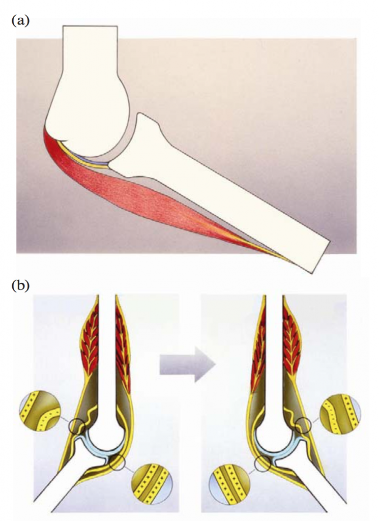
In figure 1(a), the architecture of the joint shows the muscle/tendon unit in red lying overtop, or parallel to, the ligament in yellow, which in turn is parallel to the joint capsule in blue. However, the tendon is in series with the muscle: this is the long, stringy, yellow bit at the far right of the muscle. (Note, curiously, how the top of the muscle in both (a) and (b) does not form a tendon but rather joins the bone directly! Gil Hedley calls this “bone paint.” It is as if the muscle arises directly from the bone, and it does! Sometimes, the muscle fibers connect directly with the periosteum of the bone.)
In the earlier article we learned that if the muscles are cold, engaged or generally stiffened then more of the stress created by or applied to the muscle will reach the serially aligned tissues, in this case the tendons. If the muscles are warmed up or relaxed, they will take up more of the stress and less stress will be taken by the serial tendons. In the case of parallel tissues, if the muscles are cold, tight and engaged, they will restrict the range of motion of the joint, which means that the ligaments and joint capsule are unlikely to receive any stress at all. However, if the muscles are relaxed, warm and elongated, then the ligaments may become the tissue restraining joint movement and thus will be stressed.
In short we have the following relationships:
- When muscles are cool, engaged or contracted more stress will be shared with serial tissues like the tendons, but less stress will reach the parallel tissues like ligaments and joint capsules.
- When muscles are warm, relaxed or elongated less stress will be shared with serial tissues like the tendons, but more stress will reach the parallel tissues like ligaments and joint capsules.
The Tendon Paradigm Revisited
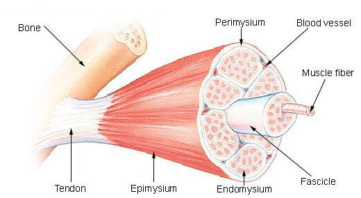
In his book Fascial Fitness, Professor Robert Schleip cites the work of Kawakami et al., who looked at the behavior of tendons during movements.[4] Many yoga teachers view tendons as being inelastic; however, tendons are quite elastic and are designed to stretch under load. (Just because they don’t stretch very much does not mean they are not elastic: it only means that they are stiff elastics.) Their ability to passively extend allows the tendons to recoil and provide energy in the opposite direction. This is the reason athletes will perform a counter-movement before an explosive leap: we squat down to jump up! The squatting down creates a stretch in the Achilles tendons and the hamstring tendons, which primes them to release this stored energy when we jump up. Kawakami illustrates this principle with a drawing, which I have modified slightly in figure 2.
In the drawing you can see that when the foot is dorsiflexed before jumping (c), both the calf muscles and the Achilles tendons are lengthened. Lengthening of the tendons creates a passive source of energy that can help us jump higher than if we used muscle effort alone. In some instances, as Robert Schleip points out, the muscles may not lengthen at all but will remain contracted and engaged. This allows even more stress to be stored in the tendons! (Remember, cold, tight or engaged muscles allow more stress into the serial tissues.) Indeed, Robert suggests that we can train the elasticity of our serial tissues by bouncing with muscles at their limit of elongation (as shown in c)) or when they are contracted and engaged.[5]
(I have heard several senior yoga teachers say that we should never passively stress our fascia and ligaments. They propose always engaging muscles to prevent stress on the fascia. However, as the above analysis shows, engaging the muscles increases stress on the serial fascial tissues, especially the tendons. So, if the intention is to avoid loading tendons, then stretches should be done with the muscles warmed up, lengthened and relaxed, not contracted. However, I do not know why this intention would arise: it is healthy to stress all tissues, including fascia. So, why try to avoid stressing our fascia by engaging our muscles? Yes, do not overstress your fascia; don’t overstress your muscles either. But do stress your tissues!)
Myofascia
The analysis so far has ignored the reality that there are fascial tissues in parallel to the muscle fibers that are neither ligaments nor joint capsules. These are the fascial wrappers that surround and invest our muscles. Indeed, about 30% of what we call a muscle is fascial tissue, which is why muscles are more accurately called myofascia. Figure 3 shows these fascial layers, which, from outer to inner, are called the epimysium, perimysium and endomysium. These fascial bags invest the muscle cells and become part of the cell. In the other direction, they expand and become the tendons and periosteum of the bones to which they connect. The fascial tissues within myofascia are in parallel to the muscle fibers; thus if our intention is to exercise or stress this fascia we could do this better if the muscle fibers are warm, relaxed and lengthened. However, the fascial expansions into tendons are in series with the muscle fibers. If we want to target them, it is better to do so when the muscles are cool, contracted and engaged.
So far, we have been looking at the classical view of joint tissues. In this paradigm, if we want to add more stress to our tendons, we should keep our muscles cool, contracted and engaged because the muscles are in series with the tendons. If we want more stress in the parallel tissues of ligaments, joint capsules and the fascia within and surrounding the muscles, we should allow the muscles to be warmer, relaxed and lengthened. But this paradigm has been called into question. There are some instances where the ligaments are not parallel to the muscles but in series with them, like tendons. In these cases, how we would stress those ligaments would be similar to how we stress tendons.
Jaap van der Wal Redefines the Ligament Paradigm
Jaap van der Wal is an anatomist who specialized in anatomy and embryology and lectured at the medical schools of the universities of Utrecht and Maastricht, in the Netherlands. In the late 1990s, he and his team pioneered a connective tissue sparing approach to dissection that didn’t sacrifice the relationships between tissues surrounding a joint. When they took more care to preserve these relationships, they discovered that our current paradigm about ligaments was rarely correct. Ligaments are not always in parallel to the muscles and tendons but rather, and quite often, are in series with them.[2]
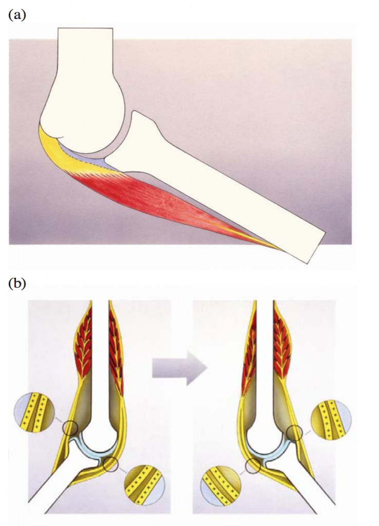
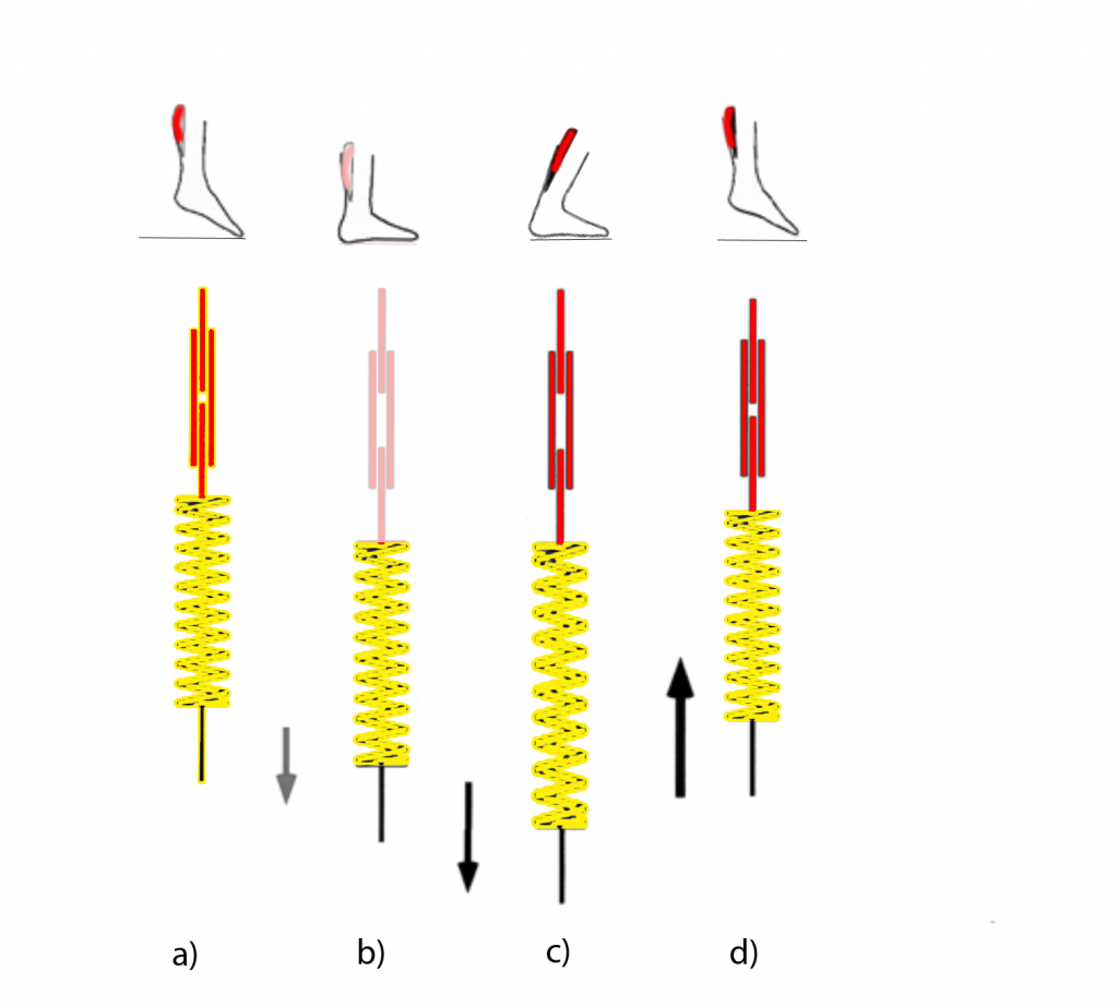
Figure 4 illustrates their findings. They discovered that ligaments do not run from bone to bone as shown in figure 1, but rather that they are in series with the muscles. They attach to the muscles and then to the bones. Their attachment to the bone is closer to the joint than tendinous attachments, so the leverage of the ligaments is smaller than that of a tendon, but clearly they are not always in parallel to the muscles as is thought in the classical paradigm. It is beyond the scope of this article to go over how Jaap and his team made this discovery. (For the full details, do check out his paper listed in footnote 2.)
Note that Jaap claims that ligaments are not always in parallel with the muscles, but there are certainly cases where they are in parallel, and the classical architecture still applies. For example, the cruciate ligaments of the knees definitely run from femur to tibia, bone to bone. And the distal (farther away) ends of the forearm muscles do form classical tendons that attach to the finger bones. But Jaap believes that the fascial tissues around the elbow joints, knees and spine follow his new paradigm rather than the classical view. (For the anatomy nerds, Jaap does not believe that the elbows’ collateral ligament complexes or even the annular ligament surrounding the head of the radius actually exist! He feels these have simply been sculpted out of the fascia that is present and do not reflect the functional reality of that fascia.)
Indeed, Jaap’s view is that there are several possible ways for bones, fascia and muscles to combine: sometimes in parallel and sometimes in series. He calls these combinations dynaments, a portmanteau of dynamic and ligaments, which dispels the passive nature of ligaments held in the classical viewpoint. Figure 5 illustrates these possibilities. This figure needs some unpacking to really understand what is going on. First, understand that there are no individual ligaments in this model, but rather dynaments that can take on many manifestations.
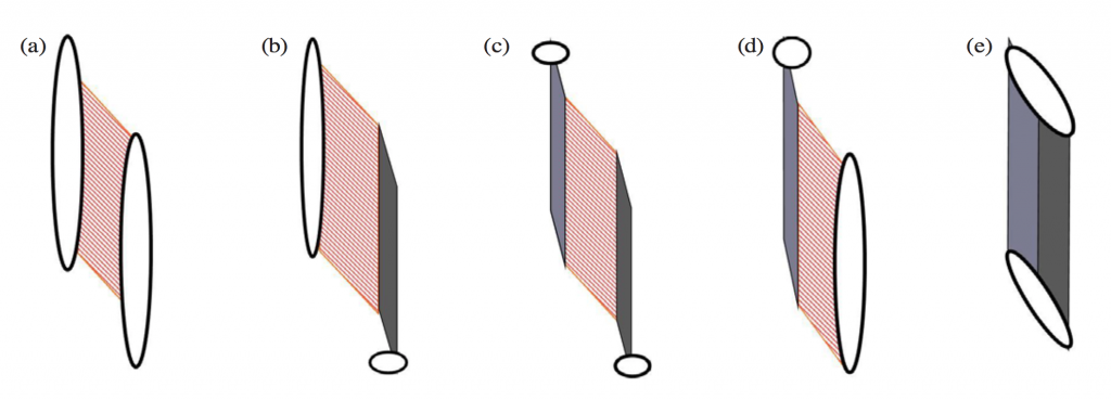
It is possible for muscles, fascia and bone to be arranged in the classical ways: part (a) illustrates the “bone paint” joining of muscle to bone. Examples include your tibialis anterior, which join directly to the tibia, or the biceps brachii with their connection to the humerus. There is no intervening fascia between them: muscle joins the bone directly. Conversely, (e) is a purely fascial connection between two bones, just as a classical ligament would suggest. This is representative of the cruciate ligaments in the knee. In (b), the muscle connects directly to the periosteum of the bone at the proximal (closer) end (as it does in (a)), but distally (farther away) the muscle becomes fascia in the form of a tendon. In (d), the reverse of (b) is shown: a muscle distally is painted onto the bones, but proximally the muscle is joined to fascial septa or aponeurosis that connects to the bone. In the middle drawing (c) we see the situation where the muscle is intermediate to the fascia and does not have a direct connection to the bones at all. Above (proximally) the muscle is joined to fascial septa or aponeurosis and below (distally) it joins fascia in the form of a tendon.[6]
In all these examples, all the muscles shown are in series with any connected fascial components. Again, there are occasions where the classical architecture is found, but Jaap believes these to be the exceptions.[7]
Implications
If we follow Jaap’s paradigm of dynaments rather than ligaments, it means acknowledging that the fascial tissues of a dynament are aligned in series with muscle tissues. Not exclusively, of course, but in general if we want to place a stress into dynament fascial tissues, which happen to be in series with our muscles, then this is best done when the muscles are engaged, contracted or cool. In the Yin Yoga style of practice where postures are held for extended periods, it is impractical to keep muscles engaged or contracted. It would be tiring and not the most efficient way to use muscles. However, we can allow the muscles to remain cool and achieve the same aim. By doing our yin asanas before the body is warmed up (before sports, exercise or more active yoga practices) the muscles will be cooler, which will allow more stress to soak into the serial fascial tissues.
Even if we choose to refuse the dynament paradigm and follow the more classical view of joint architecture, there are still fascial tissues in series with the muscles. Specifically, there are the tendons. Thus, if we want to stress the tendons in a yin-like manner, again it makes sense to do so with the muscles cooler.
However, this does not mean that there are no advantages to doing Yin Yoga when the muscles are warm. Remember, we have both serial and parallel tissues. The joint capsules and myofascia are in parallel to the muscle fibers, and if we wish to target those tissues, it makes sense to do so when the muscles are warmer, longer and more relaxed.
Should we do our Yin Yoga when the muscles are warm or cool, engaged or relaxed, longer or shorter? It depends! It depends upon which tissue you are targeting. It depends upon your intention.
FOOTNOTES
[1] See “What Are the Yin Tissues: Rethinking Elastic versus Plastic,” YinSights Newsletter (October 1, 2020). https://yinyoga.com/yin_tissues/
[2] See “Hot Yin?!” YinSights Newsletter (December 14, 2015). https://yinyoga.com/hot-yin/
[3] Jaap Van der Wal, “The architecture of the connective tissue in the musculoskeletal system—an often overlooked functional parameter as to proprioception in the locomotor apparatus,” International Journal of Therapeutic Massage and Bodywork 2.4 (December 2009).
[4] See Y. Kawakami, T. Muraoka, S. Ito, H. Kanehisa and T. Fukunaga, “In vivo muscle fibre behaviour during counter-movement exercise in humans reveals a significant role for tendon elasticity,” The Journal of Physiology 540, Pt 2 (2002): 635–646. doi:10.1113/jphysiol.2001.013459
[5] See Robert Schleip and Divo Müller, “Fascial Fitness: Fascia oriented training for bodywork and movement therapists,” Terra Rosa e-magazine 7 (August 22, 2014). https://issuu.com/terrarosa/docs/terrarosa_emag07/2
[6] For the anatomist who loves detail, here are some examples Jaap gave in his paper on which muscles exhibit these arrangements: “Distal forearm extensor carpi radialis brevis muscles and extensor digitorum muscles clearly show the (c) type of functional unit. The supinator muscle shows a variant configuration, with distal connective tissue layers that are well developed, but without an extensive connective tissue apparatus intermediating at the insertion (b). The long head of the triceps shows a similar orientation but in the “opposite direction” (d), with developed connective tissue layers proximally rather than distally. If neither a “proximal” nor a “distal” connective tissue apparatus has been developed, the result is a muscle “without aponeurosis or tendon”—for example, the [deltoid] muscle (a). If the muscular connecting and intermediating tissue has completely “disappeared,” a ligament is the consequence (e).”
[7] To quote Jaap again: “In fact, ligaments, defined as strands of RDCT [regular dense connective tissue] running from the one skeletal element to the next, are an exception. They have to be an exception. The RDCT has tissue properties of high resistance to loading, a high degree of hysteresis, and little elasticity; a ligament as interposed structure between two movable bones can therefore be constructed only if the distance between the opposite points of ligamentous insertion on the bones changes very little during the range of motion of the joint. This requires specialized design in the configuration of the bones and the joint (surfaces), and there are only a few examples of such “true” ligaments at joints in the body: cruciate ligaments of the knee joint or ligamentum apicis dentis in the atlanto-occipital joint.”